GEHC Revolution Maxima Product Web Page Pre Scan Image 1

Right place, less time

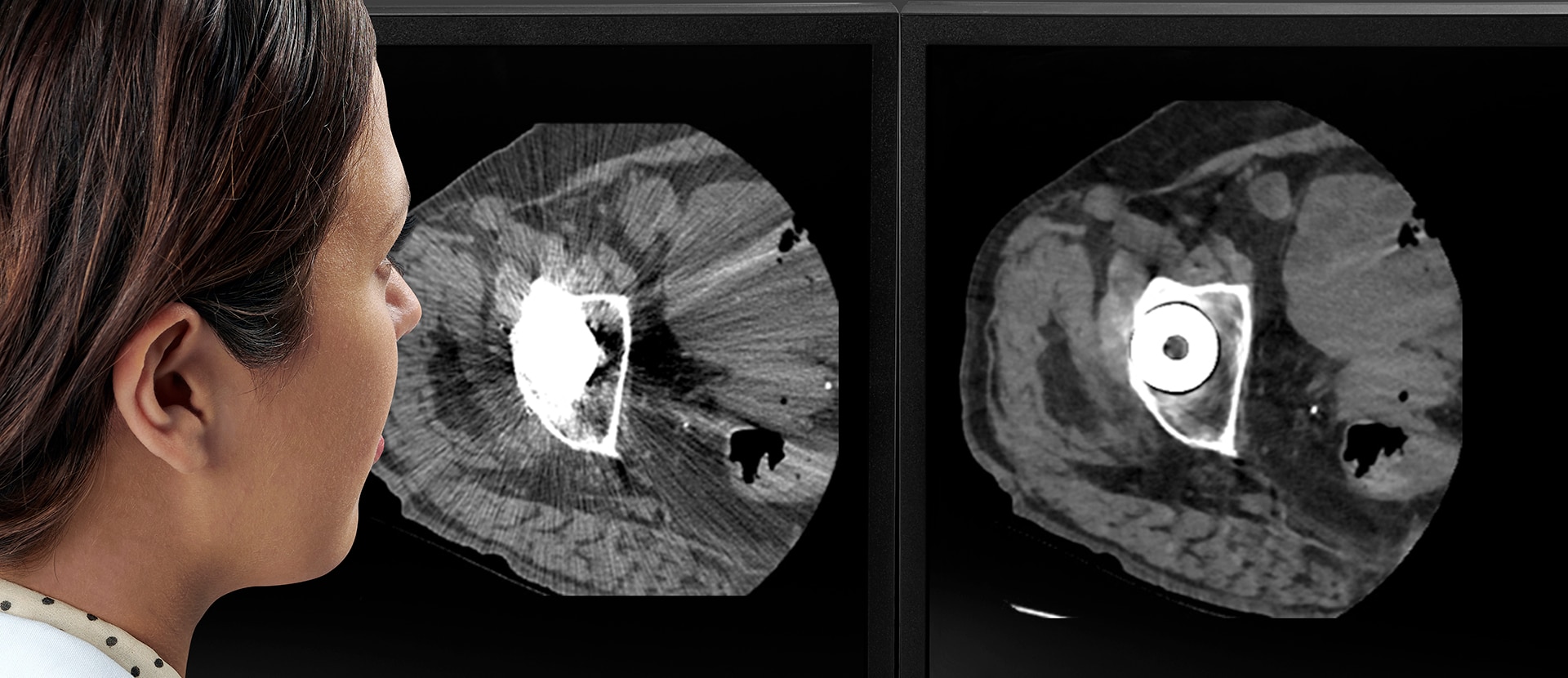
No matter how you look at it, all patients are at risk of being mispositioned. When a patient is mispositioned, it may lead to up to a 38 percent increase in dose1 and up to a 22 percent increase in image noise2 . Positioning challenges are difficult to resolve because they can stem from a combination of variables, including the inherent inconsistency of a manual workflow, patient discomfort and technologist experience.

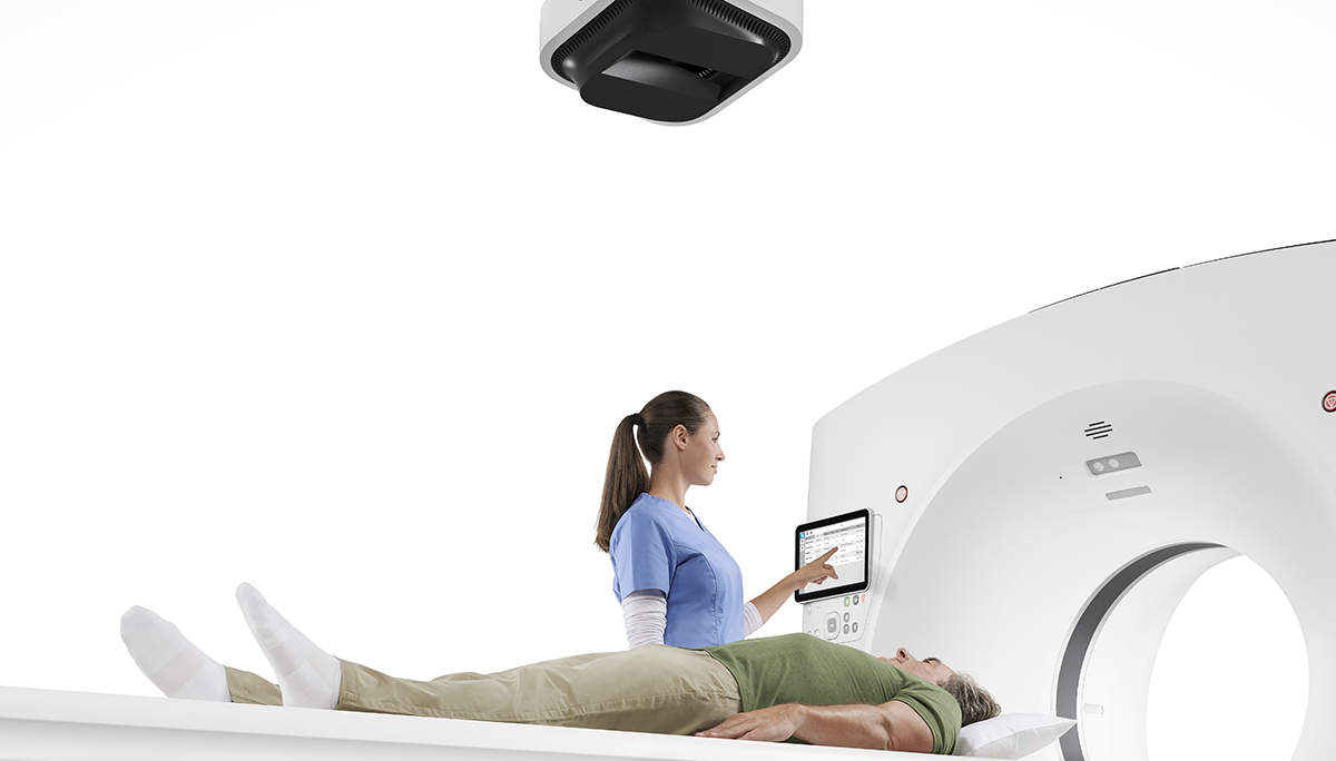
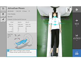
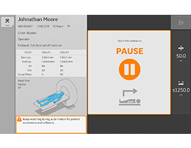
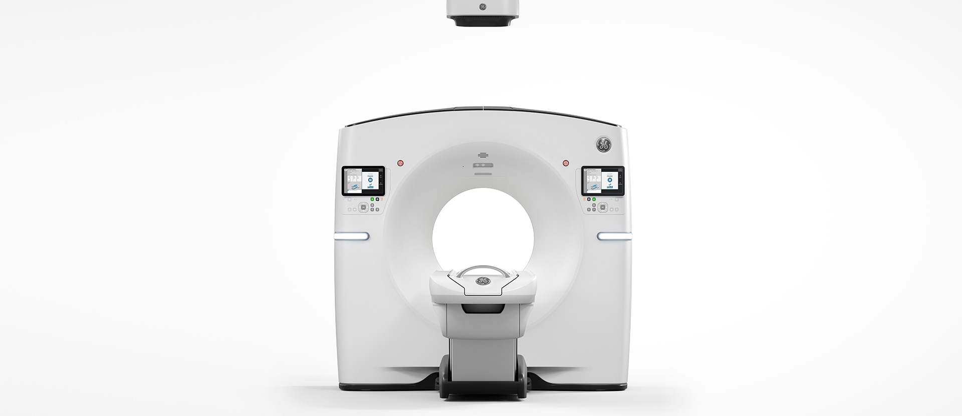
Our innovative AI-based Auto Positioning* completely automates this step. The Xtream camera uses real-time depth sensing technology to generate a 3D model of your patient’s body. Then, using our deep learning algorithm, Revolution Maxima pinpoints the 3D center of the scan range and automatically aligns it with the isocenter of the bore. With one click, Auto Positioning uses all of this information to automatically center your patient for a completely hands-free positioning experience.

Auto Positioning streamlines your patient setup and, most important of all, frees up your technologists so they can focus on making your patients feel more comfortable.

*Auto Positioning is 510(k) pending at FDA. Not available for sale in the United States.


One-click, hands-free automation

Revolution Maxima simplifies the entire patient setup process. Innovative auto centering technology is at the core of our improved scan experience, but it starts with related protocol recommendations. By comparing the exam description against a database of scan protocols, the system displays a short list of protocols to choose from. What used to take valuable time searching for the right protocol and then manually positioning the patient in the bore, can now be done with a quick selection and the simple click of a button.


Smart select

Quickly get to the appropriate protocol by choosing from a simple list of related protocols.


Auto center

By calculating the 3D center of the scan range, the system knows exactly how to align the table in the bore.


Click to position

With the click of a button, automatically position your patient at the start location of the scan.

GEHC Revolution Maxima Product Web Page During Scan Image 1

It comes with everything you need

We wanted to provide you with one CT that comes preloaded with all of the capabilities essential to getting the right information at the right image quality across a diverse range of clinical needs. Built around our Clarity imaging chain, Revolution Maxima provides 0.28 mm3 spatial resolution and makes 40 mm imaging routine.

Our leading dose reduction software, ASiR-V™, is also available. This advanced noise and object modeling reconstruction software allows you to consistently image with up to 82 percent4 less dose compared to filtered back projection image reconstruction. Together, these core technologies provide you with everything you need to get the right diagnosis, the first time, at the lowest dose possible.

GEHC Revolution Maxima Product Web Page Post Scan Image 1

Findings you can clearly dictate

Your CT experience doesn’t end with the generation of a high-quality image. You also need intelligent applications that continue to support you once those images reach the reading room. We’ve developed a collection of smart applications for cardiac, stroke, oncology and metal artifacts that do just that.


Smart MAR

Single acquisition metal artifact reduction.


Smart Cardiac

Set up complex cardiac procedures quickly, reliably and repeatedly.


Smart Stroke

Perform stroke assessment scans with perfusion shuttle technology and assess patient status quickly and accurately.

One great image after another

Great imaging performance is more than just a beautiful clinical image. For high-throughput environments, how you get to the image is just as important as the image itself. And every step in the process matters. You need to consider how quickly a scan can be set up, how easily your applications can be applied during the scan and how effectively you can produce a report from the images you acquire. All of this goes into the great imaging performance you can expect with Revolution Maxima.

GEHC Revolution Maxima Product Web Page Services Image 1

A CT that continuously works behind the scenes

When it comes to the operational and financial success of your imaging department, there is a lot your CT can do for you in the background using remote diagnostics, predictive analytics, cloud-based solutions and subscription-based services. Take Tube Watch as an example. Through a combination of artificial intelligence, machine learning and software analytics called Digital Twin technology it creates a customized, digital model of your tube, system and usage profile. Using that digital model, Tube Watch can predict when your tube is going to need to be replaced. This is just one of many ways Revolution Maxima is maximizing your capabilities.

A CT that can do it all

As a high-performing imaging department, you don’t need a range of CT options to pick and choose which capabilities you need. From scan setup, to image acquisition, to the final report, there is a lot that goes into high performance CT imaging. What you need is a system that can do it all.

Revolution Maxima provides innovative solutions for your entire CT experience from referral to report, including an incredible new way to automate patient positioning. Beyond that, it’s a scalable, upgradable system that leaves the door open to grow with your practice.

  1. Kaasalainen, T., Palmu, K., Reijonen, V., & Kortesniemi, M. (2014). Effect of patient centering on patient dose and image noise in chest CT. American journal of roentgenology, 203(1), 123-130.
  2. Toth T, Ge Z, Daly MP. The influence of patient centering on CT dose and image noise. Med Phys 2007; 34:3093–3101.
  3. Calculated based on MTF 4% value in X/Y. 4% MTF is measured under 120kv, 200mA, 1.0sec gantry rotation and Edge Plus kernel.
  4. In clinical practice, the use of ASiR-V may reduce CT patient dose depending on the clinical task, patient size, anatomical location, and clinical practice. A consultation with a radiologist and a physicist should be made to determine the appropriate dose to obtain diagnostic image quality for the particular clinical task.

JB00223SG