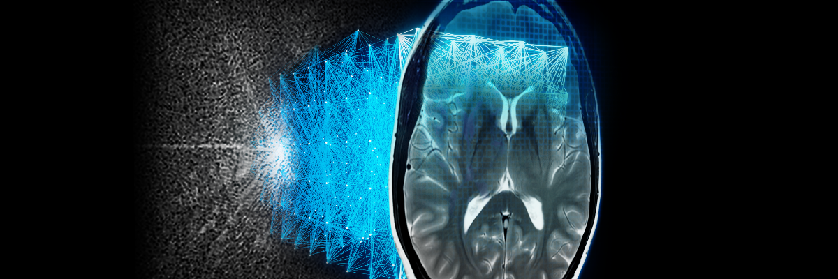
Artificial intelligence (AI) applications in the field of medical imaging are no longer just conceptual. They are currently proving their value in clinical practice, offering clinicians assistance, for example, in triaging and prioritizing urgent cases in X-Ray, using deep learning to improve the quality of low-dose CT scans, and also in applying deep‐learning models for image reconstruction in MRI.
A recent webinar hosted by GE Healthcare brought together a panel of clinicians to discuss some of the ways they’re seeing these AI solutions impact MR cases from a clinical standpoint, as well as improvements they’re seeing in productivity and return on investment (ROI) that impact their overall imaging operations and workflow.
Deep-Learning MR Image Reconstruction in Clinical Practice
Applying AI and deep-learning algorithms to MR image reconstruction is an exciting realization of technology that is enabling improvements in MR that haven’t been possible using traditional reconstruction methods. Healthcare providers are using this technology to produce high-quality images with shorter scan times, overcoming the historical trade-offs in MR between scan time and image quality.
Expert panelists on the webinar, “Intelligently Efficient—Brought to You by AIRTM Recon DL” are all currently using GE Healthcare’s AIR™ Recon DL* technology, which is a deep-learning trained algorithm to reconstruct sharper images that leverages all of the raw acquisition data, delivers a higher signal-to-noise (SNR) ratio with user-selectable improvement levels, and is compatible with all anatomies.
Melanie Atkins, MD, a radiologist with Fairfax Radiology Centers in Fairfax, Virginia, presented clinical case studies illustrating the image quality and resolution improvements she’s been able to achieve in her clinical practice using AIR™ Recon DL. She presented clinical cases of MR imaging for cervical cancer, as well as MR cholangiopancreatography (MRCP), a non-invasive imaging technique used to evaluate the liver, gallbladder, bile ducts, pancreas and pancreatic duct for disease. Dr. Atkins was able to shorten the traditionally longer acquisition times for her patients’ pelvic and abdominal exams, which often cause organ blur.
In study after study, Dr. Atkins noted that her patients, some of whom are elderly and struggle in the MR, are spending less time in acquisition, and she is getting far superior image quality to inform clinical decisions on diagnosis and treatment. Her colleagues have also embraced the use of AIR™ Recon DL for all musculoskeletal and neuroimaging based on her clinical results.
Protocol Optimization for Every Imaging Scenario
As Dr. Atkins demonstrated, the application of deep-learning reconstruction to traditional MR imaging has resulted in superior image quality while reducing patient’s time in the scanner. Christopher Ahlers, MD, next noted that these types of clinical results are not only possible, as Dr. Atkins illustrated, but also extremely easy to use and this AI solution can be incorporated seamlessly into clinical practice without additional work for the radiologist. Dr. Ahlers, a radiologist and Managing Partner at radiomed Practice for Radiology and Nuclear Medicine in Wiesbaden, Germany, presented cases using deep-learning reconstruction to control the output he was looking for in each case.
"...We can drastically increase the contrast-to-noise ratio, which really improves lesion conspicuity. It allows us to push protocols or applications to levels that would otherwise be incompatible with conventional reconstruction and works in any anatomy.”
“Mostly, it’s about protocol optimization,” Dr. Ahlers explained. “When we optimize the original sequence to increase spatial resolution or improve scan time, we are likely seeing some noise on the images. This noise can be removed by using AIR™ Recon DL, in order to regain signal we lost, or even improved SNR. It is easy to switch on AIR™ Recon DL, you just select preferred SNR level from the drop-down menu, you can even choose the original reconstruction. Our observation is that we can drastically increase the contrast-to-noise ratio, which really improves lesion conspicuity. It allows us to push protocols or applications to levels that would otherwise be incompatible with conventional reconstruction and works in any anatomy.”
Dr. Ahlers also noted several new opportunities for imaging that are enabled by deep- learning image reconstruction, such as cardiac myocardial perfusion imaging, where he’s been able to increase the spatial resolution in order to decrease diaphragm artifacts, as well as free-breathing imaging sequences, which require virtually no cooperation from the patient and achieve great image quality with low noise levels.
Intelligently Efficient Changes in MR Workflow Productivity
The clinical images presented by the participating clinicians on the webinar illustrated the dynamic improvements in image quality achievable as a result of using deep-learning image reconstruction, but two other clinicians shared the equally impressive quantitative performance results they’ve achieved by incorporating deep-learning reconstruction into their MR workflows.
Lawrence Tanenbaum, MD, Vice President and Medical Director, MRI, CT and Advanced Imaging for Radnet, Inc., in New York City, is using deep-learning reconstruction at one of their busiest centers in Manhattan.
"Across the board, and across the range of exams, we run anywhere between 30-50% reduction [in scan times]."
“Initially, you think, this will help me go faster, and it does, but it’s actually going to elevate your entire imaging chain because there’s such powerful noise reduction and greater efficiency. Across the board, and across the range of exams, we run anywhere between thirty and fifty percent reduction [in scan times]. We originally had 20-minute appointment slots; now they are dropping to 15-minute slots. With this innovation, we can have one more patient an hour. That’s about ten more patients a day. And frankly, aside from the throughput benefits, there is improved operational consistency. It allows us to stay on schedule. It gives us a greater ability to accommodate add-ons and handle the typical disruptions that will always come up like insurance certifications and implanted devices. It certainly keeps us on schedule and allows us to reduce our overtime needs,” Dr. Tanenbaum explained.
Dr. Ahlers agreed, adding that beyond the improved quality of the images that allows the radiologist easier depiction of lesions and lesion conspicuity, it’s causing them less eye fatigue because they are easier to read.
“I personally find it less exhausting,” Dr. Ahlers explained. “It’s just easier to read through these beautiful images.”
Reduction in MR Scan Time and the Impacts on ROI
Randall Stenoien, MD, offered a perspective from a private practice of radiologists. As owner and CEO of Houston Medical Imaging, Dr. Stenoien commented that investing in new technology, especially expensive technology, needs to be very thoughtful. It’s a business decision.
“I found that in my practice,” explained Dr. Stenoien, “the secret to my financial survival has been the fact that we’re able to do more complex exams. There is more reimbursement there. The downside is that these exams are substantially more time-intensive and the scheduling slots need to be much longer.”
Dr. Stenoien made the decision to invest in opening a new imaging center in October 2019 with a twelve-month timeline to profitability based on revenue projections. Unfortunately, because of the COVID-19 pandemic, he was forced to close the center in March 2020, his plan to profitability dashed. When Dr. Stenoien heard about the potential of deep-learning reconstruction in MRI with AIR™ Recon DL to accelerate image acquisition, he didn’t need much more convincing, but then he saw the image quality.
“The image quality was fantastic immediately,” he explained. “And we were seeing dramatic reduction in scan times. One of our first patients was a pituitary scan; 30 minutes 40 seconds. In January on the same scanner, the scan was 14 minutes and 42 seconds, a 52 percent reduction. And when I sent those images to my neuroradiologist, I immediately got a phone call to ask what in the world had happened. It was the prettiest pituitary scan she’d ever seen.”
Dr. Stenoien commented that the learning curve to adopt AIR™ Recon DL was so rapid that his team was able to immediately adopt their protocols and increase their patient load by 50 percent and he spoke about the financial impact of his investment, especially considering the global pandemic and the additional time that will be needed to incorporate cleaning protocols in between patients.
“How did this affect our profitability? To me, that’s the most compelling part of the story. We went live at the end of September [2020] and prior to going live, we were doing on average 10-12 patients a day. With AIR™ Recon DL, we were able to add four time slots a day on average, and the reimbursement data, it’s not linear. October of 2020 was the first month we went into the black. We not only broke even, but we were able to make some money and see a return on investment. As we come out of COVID and increase volumes further, we’re going to have a really tremendous opportunity to be profitable. It’s been a wonderful experience,” Dr. Stenoien concluded.
To hear more from these four clinicians and their experiences with AIR™ Recon DL, as well as to see the compelling images enabled by AIR™ Recon DL, visit the GE Healthcare Experience and access the webinar in the Innovation Theater.
To learn more about AIR™ Recon DL, please visit AIR™ Image Quality | GE Healthcare.
*Not yet CE marked for 1.5T. Not available for sale in all regions.
Also available on YouTube for viewing.


