Medical imaging continues to grow and X-ray technology remains one of the fundamental types of diagnostic imaging. X-ray contributed to more than 80 percent of the estimated 4.2 billion imaging procedures that were performed in 2019.[1] Some of the key attributes that enable imaging a high volume of patients is that it is noninvasive and fast. The most recent innovations in X-ray not only focus on incorporating advanced artificial intelligence (AI) tools to improve operational and reading workflows, but also on improving the ergonomics of these systems to reduce the stress and strain on staff.
As a leader in the industry, GE Healthcare understands these demanding aspects of the technologists’ workflow and has focused new X-ray system designs on ergonomics, usability and using AI to reduce the cognitive workload for technologists and radiologists. Advances in X-ray technology, and adoption of AI tools to streamline workflow have made significant impacts on the delivery of care in X-ray, especially during the challenges presented by the global pandemic.
“The design objective for our new portable and fixed x-ray systems were focused on creating an effortless workflow for the technologists, using embedded AI technologies to improve clinical excellence, as well as constructing the systems with rugged reliability for our customers, so they can focus on what matters most, delivering care to each patient,” said Andrew DeLaO, Chief Marketing Officer, Women’s Health and X-ray at GE Healthcare.
Advances in X-ray technology address physical and cognitive workload challenges
Radiologic technologists are essential members of the health care team. The work they do, both with the patients and behind the scenes impacts patient outcomes and the operational success of the radiology department. Unfortunately, more than 70 percent of radiologic technologists will experience work-related stress or strain at some point in their career.[2]
X-ray technologists are often one of the first to encounter a patient, who has abnormal symptoms and the ED or their physician needs more information. Diagnostic X-ray exams require technologists to lift, transfer or move heavy objects many times within each shift.[3] Working with older equipment, technologists often need to complete numerous workflow steps and complicated patient set-ups with limited clinical applications embedded on the systems.
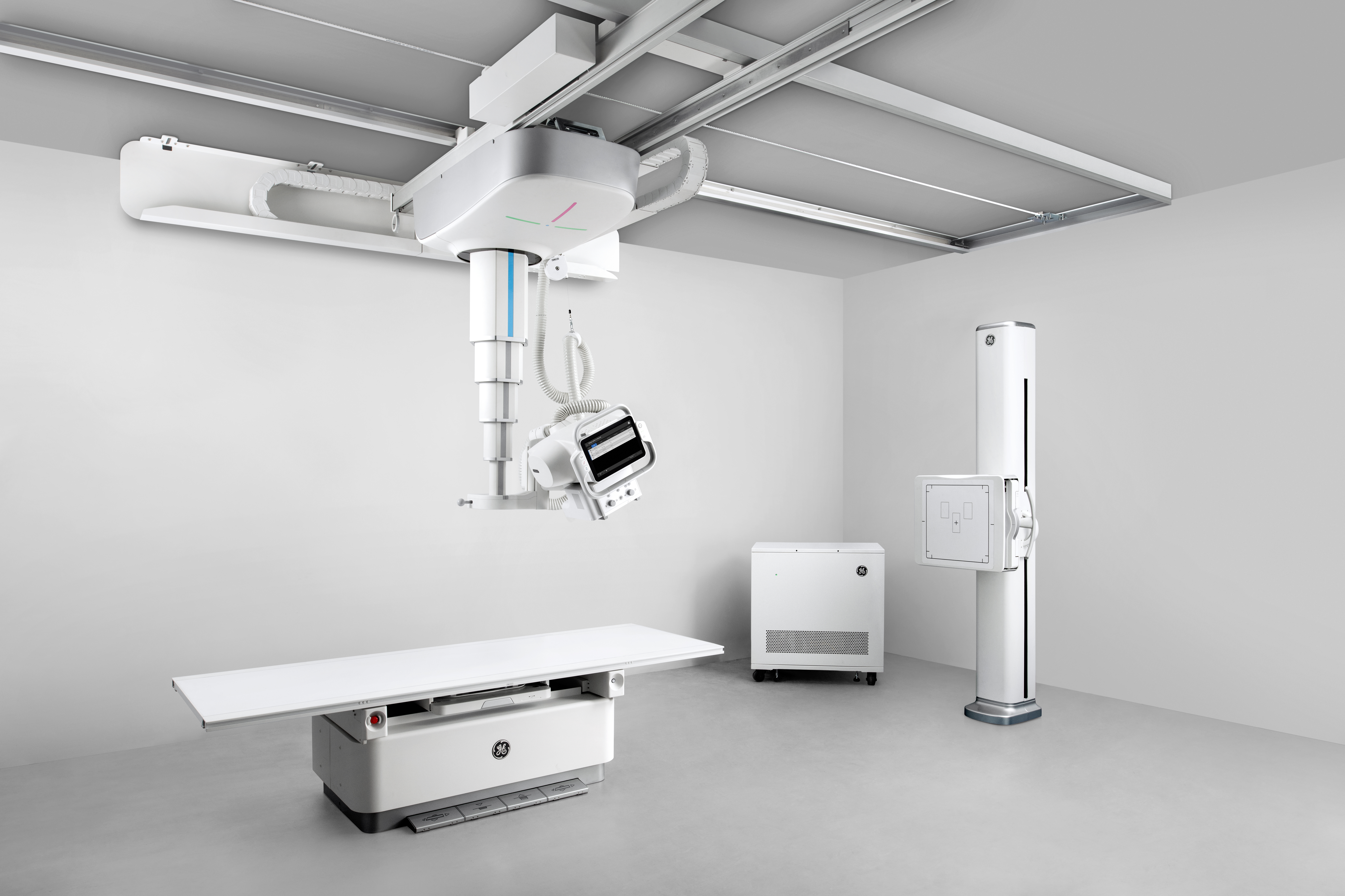
For both of GE Healthcare’s new mobile and fixed X-ray systems, features such as motorized assist and auto positioning remove the physically demanding aspects of the clinical workflow, while its effortless workflow capability reduces the technologists’ cognitive workload.
The systems’ automated tools are intended to assist technologists with correct anatomical positioning and confirm protocol selections at the time of the scan. It also enables images to auto rotate to save time for reading radiologists.
“Prior to the [new fixed X-ray] Definium™ Tempo,” explained Jeannie Miller, Associate Director of Radiology at North Central Bronx Hospital in New York, “everything was done manually — loading a cassette into the bucky, aligning your overhead tube with your table, aligning it back to the wall. It creates inconsistent imaging. Now, one button aligns the overhead bucky with the table and the wall. There is no more physical labor…Auto positioning has taken all of that away. [Technologists] are willing to do overtime now because there is less strain on the actual body.”
Improving clinical workflow without disruption
The development of AI-based applications in healthcare continues to grow[4] but oftentimes, the options may be overwhelming or can take time for the implementation and integration into the clinical workflow instead of impacting clinical care quickly.
Industry leaders, like GE Healthcare are focused on developing AI and advanced clinical tools and enabling easy and quick implementation of these tool. The goal is to increase efficiencies across the entire radiology workflow without increasing the administrative and training burden on radiologists and technologists.
Amit Gupta, MD, Modality Director of Diagnostic Radiography at University Hospital Cleveland Medical Center and Assistant Professor of Radiology at Case Western Reserve University, Cleveland is a pioneer in implementing new artificial intelligence tools into the existing clinical workflow for maximum impact. Working collaboratively with GE Healthcare, he participated in the development of their collection of on-device AI algorithms for X-ray, the Critical Care Suite.
Dr. Gupta recently created and published a paper outlining the institutional roadmap for the successful implementation of new AI tools, highlighting the milestones in the end-to-end integration of the tool into clinical practice.[5]
“A lot of work has been done on the side of developing the AI algorithm, but the clinical implementation remains relatively low,” Dr. Gupta explained. “Hand in hand with the high accuracy of the Critical Care Suite algorithm, is its ability to seamlessly integrate into your existing workflow.”
Artificial intelligence and X-ray — impacting patient care with speed and efficiency
AI tools have proven to be especially helpful in triaging patients and highlighting cases that require the most urgent attention. To improve triage of urgent cases, Critical Care Suite automatically analyzes images, upon acquisition, for critical findings. Triage notifications are sent directly to PACS and flagged for prioritized radiologist review.
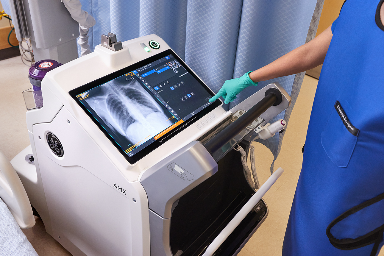
Embedded in both fixed and mobile X-ray systems, the AI tool streamlines the workflow with features such as automatic image rotation, quality check, and an intelligent protocol check to detect errors during the acquisition, allowing the technologist to make a correction before the patient leaves the exam.
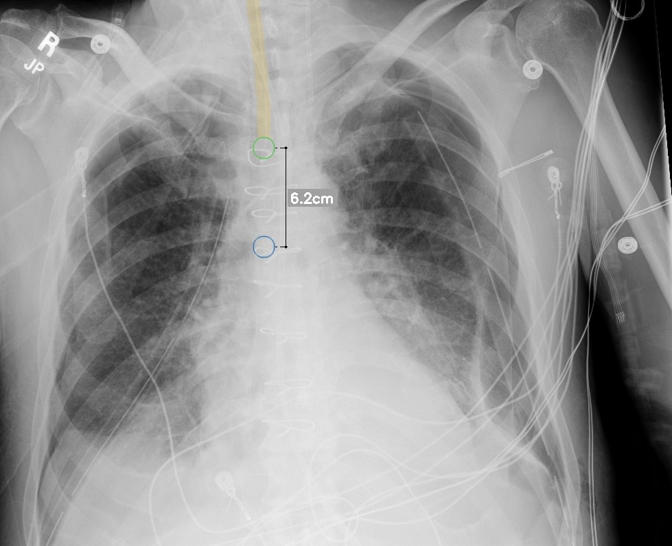
The AI algorithms detect critical findings using deep learning, trained from thousands of chest X-ray images. They also provide automated measurement that help with the accurate placement of endotracheal (ET) tubes. Dr. Gupta and his team are using the AI Critical Care Suite to verify proper ET tube placement, triage urgent cases, and detect the presence of pneumothorax.
“Seconds and minutes matter when dealing with a collapsed lung or assessing endotracheal tube positioning in a critically ill patient,” explained Dr. Gupta. “In several COVID-19 patient cases, the pneumothorax AI algorithm has proved prophetic – accurately identifying pneumothoraxes/ barotrauma in intubated COVID-19 patients, flagging them to radiologist and radiology residents, and enabling expedited patient treatment. Altogether, this technology is a game changer, helping us operate more efficiently as a practice, without compromising diagnostic precision.” [6]
Building a world that works for X-ray
Advanced digital X-ray technology and AI tools that automate tasks and provide clinical decision support are having a significant impact on radiology today and will continue to do so into the future. These paradigm shifts are enabling technologists and radiologists to complete exams with accuracy and precision, with less physical demands, and more time to focus on the patient. The increasing adoption and use of AI and machine learning in the imaging space will continue to improve and streamline workflow, addressing today’s challenges, and whatever challenges may be on the horizon.
RELATED CONTENT
X-ray spotlights from GE Innovation Theater
- Definium™ Tempo. Definitive insights, exceptional X-ray experience
- Introducing Definium™ Tempo, a highly automated, AI-powered digital X-ray imaging solution equipped with intelligent applications, allowing technologists to focus on what matters most - their patient.
- View replay now
- Clinical deployment of Critical Care Suite 2.0: On-device X-ray AI helping assess endotracheal tube placement
- As many as 25% of intubations outside of the operating room result in mispositioned ET Tubes, which can lead to severe complications. Dr. Amit Gupta shares UH Cleveland’s experience with integrating the Critical Care Suite in daily clinical practice: an on-device X-ray AI that helps assess ET Tube positioning.
- View replay now
- Innovation in motion with Mobile X-ray, AMX™ Navigate
- Designed to address the stress and strain in positioning for imaging, AMX™ Navigate, our latest digital mobile X-ray system, provides a new level of clinical excellence with AI clinical decision and effortless workflow support.
- View replay now
Not all products or features are available in all geographies. Check with your local GE Healthcare representative for availability in your country.
[1] 2019 Global Imaging Outlook Report, IMV, https://imvinfo.com/
[2] https://journals.sagepub.com/doi/pdf/10.1177/8756479315580020
[3] Elshami W,Abuzaid M, Ateeq E, Al Fozan S, Bensahraoui N, Zira D. Prevalence of repetitive stress injuries among radiologic technologists. Am J Diag Imaging. 2018;3(1):12-17.
[4] https://www.pharmaceutical-technology.com/features/ai-in-healthcare-2021/
[5] https://www.jacr.org/article/S1546-1440(21)00741-9/pdf
[6] https://www.businesswire.com/news/home/20201123005895/en/GE-Healthcare-Announces-First-X-ray-AI-to-Help-Assess-Endotracheal-Tube-Placement-for-COVID-19-Patients


