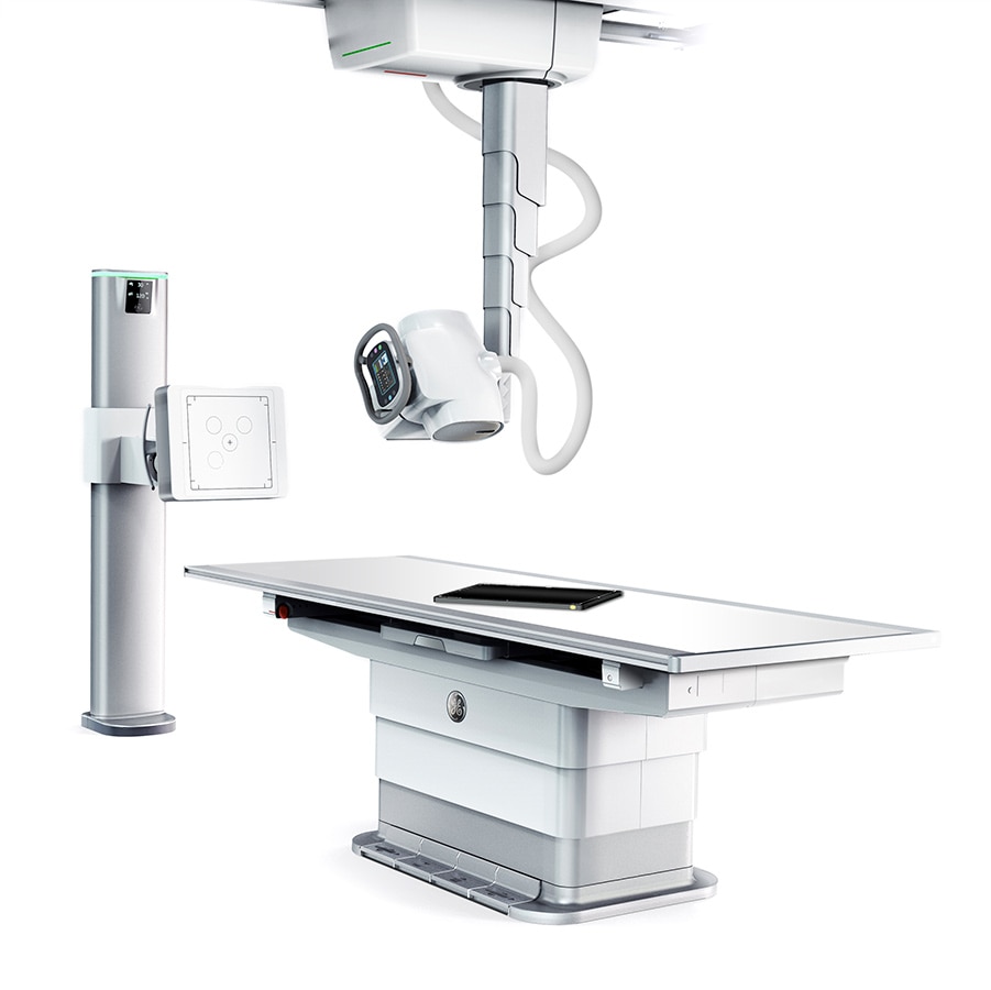
A new frontier in digital X-ray image quality. Effortless precision.
With Discovery XR656 HD, no more unnecessary X-ray image adjustments and repeated exams. Just sharp detail and the balanced contrast and brightness you need at low dose, right from that very first image
Get the diagnostic clarity you need from that first X-ray
$name
Extraordinary anatomical detail at low dose in every X-ray image.
Helix™ advanced image processing algorithms harness the full high-resolution power and exceptional dose efficiency of FlashPad HD detectors to deliver outstanding clarity and extraordinary anatomical detail where it matters most.
Up to 40% improvement in detectability of fine structures1
The power of Helix™ advanced image processing coupled with FlashPad HD improves small detail detectability by up to 40%1 thanks to ultra-high resolution and enhanced noise control.
$name
Consistent brightness and contrast
Helix delivers consistent brightness and contrast across variations in dose exposure with Smart Windowing and enhanced Contrast Restoration.
Quadruple your resolution
The FlashPad HD detectors pack four times more pixels per area for sharp X-ray images, plus they capture extraordinary anatomical detail at low dose. Available in 10 in x 12 in & 14 in x 17 standard cassette sizes.
$name
Exceptional dose efficiency for your tiniest patients (and the large ones too)
The ultra-high dose efficiency helps enhance diagnostic imaging quality at low dose for all patient types.
Excellent handling of metal implants
Clear bone-metal interface without halo artifact.
Improving Patient Experience and Workflow. AutoRAD Comprehensive Workflow Automation Suite.
Auto Field-of-View
Predefined collimation sizes for each view.Auto-tracking
Maintain SID &tube-detector alignment with table and wall stand receptor automatically.QuickCharge
Detector charging in the table and wall stand bucky.New User Interface
Redesigned navigation and quick tools for fewer clicks and intuitive operationQuickCharge
Detector charging in the table and wall stand bucky.QuickShare
Hassle-free sharing and pairing of multiple wireless detectors.Auto Protocol Assist
Automatic selection of anatomy & technique based on modality work list.QuickConnect
Automatic wifi channel switching to avoid wireless interference.
Your patient’s safety, comfort and dignity in mind.
A bariatric X-ray table capable of supporting up to a 400 kg / 882 lbs3
that lowers to 50cm / 20 inches
Data isn’t just about looking backwards. It helps you plan the future.
Ready when you are
Supporting Materials
You may also be interested in
1. Source: GE whitepaper : High resolution for improved visualization (DOC2045904)
2. "A paediatric X-ray exposurechart"; Stephen P Knight; Journal of Medical Radiation Sciences, 2014
3. Table weight limit: 400kg/882 lb static and 320kg/705 lb dynamic (elevating)
4. Service and education offers may vary by country, check with your local representative